


Oct 02, 2006
Web Journals Take On Peer Review
LOS ANGELES - Scientists frustrated by the iron grip that academic journals hold over their research can now pursue another path to fame by taking their research straight to the public online.
Instead of having a group of hand-picked scholars review research in secret before publication, a growing number of internet-based journals are publishing studies with little or no scrutiny by the authors' peers. It's then up to rank-and-file researchers to debate the value of the work in cyberspace.
The web journals are threatening to turn the traditional peer-review system on its head. Peer review for decades has been the established way to pick apart research before it's made public.
Next month, the San Francisco-based nonprofit Public Library of Science will launch its first open peer-reviewed journal called PLoS ONE, focusing on science and medicine. Like its sister publications, it will make research articles available for free online by charging authors to publish.
Read the full story
17:03 Posted in Research tools | Permalink | Comments (0) | Tags: research tools
Sep 30, 2006
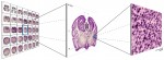
Endoscopic eye tracking system for fMRI
Endoscopic eye tracking system for fMRI.
J Neurosci Methods. 2006 Sep 13;
Authors: Kanowski M, Rieger JW, Noesselt T, Tempelmann C, Hinrichs H
Here we introduce a new video-based real-time eye tracking system suitable for functional magnetic resonance imaging (fMRI) application. The described system monitors the subject's eye, which is illuminated with infrared light, directly at the headcoil using an endoscopic fibre optical system. This endoscopic technique assures reliable, easy-to-use and fast adjustment. It requires only a minimal amount of equipment at the headcoil and inside the examination room. Moreover, the short distance between the image acquisition optics and the eye provides high spatial tracking resolution. Interference from physiological head movement is effectively reduced by simultaneous tracking of both eye and head movements.
19:25 Posted in Neurotechnology & neuroinformatics, Research tools | Permalink | Comments (0) | Tags: neuroinformatics, eye-tracking
Sep 19, 2006
Using VR to assess neglect
Pseudoneglect in back space.
Brain Cogn. 2006 Aug 23;
Authors: Cocchini G, Watling R, Sala SD, Jansari A
Successful interaction with the environment depends upon our ability to retain and update visuo-spatial information of both front and back egocentric space. Several studies have observed that healthy people tend to show a displacement of the egocentric frame of reference towards the left. However representation of space behind us (back space) has never been systematically investigated in healthy people. In this study, by means of a novel visual imagery task performed within a virtual reality environment, we found that representation of right back space is perceived as smaller than the left. These results suggest that there is a selective compression or distortion for mental representation related to the right space behind us.
17:35 Posted in Research tools, Virtual worlds | Permalink | Comments (0) | Tags: virtual reality, cybertherapy
Sep 18, 2006
VR experiment casts light on anxiety
Context Conditioning and Behavioral Avoidance in a Virtual Reality Environment: Effect of Predictability.
Biol Psychiatry. 2006 Aug 31;
Authors: Grillon C, Baas JM, Cornwell B, Johnson L
BACKGROUND: Sustained anxiety can be modeled using context conditioning, which can be studied in a virtual reality environment. Unpredictable stressors increase context conditioning in animals. This study examined context conditioning to predictable and unpredictable shocks in humans using behavioral avoidance, potentiated startle, and subjective reports of anxiety. METHODS: Subjects were guided through three virtual rooms (no-shock, predictable, unpredictable contexts). Eight-sec duration colored lights served as conditioned stimuli (CS). During acquisition, no shock was administered in the no-shock context. Shocks were paired with the CS in the predictable context and were administered randomly in the unpredictable context. No shock was administered during extinction. Startle stimuli were delivered during CS and between CS to assess cued and context conditioning, respectively. To assess avoidance, subjects freely navigated into two of the three contexts to retrieve money. RESULTS: Startle between CS was potentiated in the unpredictable context compared to the two other contexts. Following acquisition, subjects showed a strong preference for the no-shock context and avoidance of the unpredictable context. CONCLUSIONS: Consistent with animal data, context conditioning is increased by unpredictability. These data support virtual reality as a tool to extend research on physiological and behavioral signs of fear and anxiety in humans.
17:30 Posted in Research tools, Virtual worlds | Permalink | Comments (0) | Tags: virtual reality, cybertherapy
Relevant psychology journals in new media/communication technology
The website of University of Twente (faculty of Behavioral Sciences) has a list of relevant journals in HC/ergonomics; ISI impact factors is also provided for most of them.
The complete list can be accessed here
12:54 Posted in Call for papers, Research tools | Permalink | Comments (0) | Tags: research tools, human-computer interaction
Sep 10, 2006
Portable MRI
Via the Neurophilosopher's blog
Alexander Pines and his colleagues at the U.S. Department of Energy’s Lawrence Berkeley National Laboratory are working on a new laser-based MRI technique which may lead to the development of a cheap and compact scanning device.
The experimental technique is based on a method called atomic magnetometry, which allows to detect the magnetic signals produced by water molecules without the large magnets or complex cooling systems used in conventional fMRI.
From the LBNL website:
Alexander Pines and colleagues at Berkeley Lab have developed a method to improve NMR/MRI resolution either inside of poorly shimmed magnets or outside of portable one-sided magnet systems, which accommodate arbitrarily sized samples. This technique will enable for the first time the collection of multidimensional NMR/ MRI information in cases where on-the-spot medical diagnosis is critical, where samples cannot be moved to or placed inside of a superconducting magnet, or where inexpensive, highly inhomogeneous magnets are being used. Other ex situ systems give relaxation data and sometimes slice-selective images, but not spectra and true 3D images.

21:35 Posted in Neurotechnology & neuroinformatics, Research tools | Permalink | Comments (0) | Tags: research tools
Nanowires Listen In on Neurons
Via Neuroguy
MIT’s Technology Review has an interesting article that describes the development of silicon nanowires to measure small electrical signals on the same neuron:
The research group, led by Charles Lieber, professor of chemistry at Harvard University, has developed techniques for synthesizing large arrays of silicon nanowires, which act as transistors, amplifying very small electrical signals from as many as 50 places on a single neuron. In contrast, the most precise existing methods can pick up only one or two signals from a neuron. By detecting electrical activity in many places along a neuron, the researchers can watch how it processes and acts on incoming signals from other cells.The nanowires are about the same size as the branches that neurons use to communicate with one another. William Ditto, professor of biomedical engineering at the University of Florida, says neurons probably send the same kinds of signals to the nanowires as they do to other neurons. As a result, the nanowires could provide a realistic view of a neuron’s complex firing patterns.
21:21 Posted in Neurotechnology & neuroinformatics, Research tools | Permalink | Comments (0) | Tags: neurotechnology
Sep 08, 2006
Visualization of literature trends
Via infosthetics

CiteSpace is a network data visualization technique that facilitates the detection of emerging trends and transient patterns in scientific literature.
From Infoesthetics:
CiteSpace is based on 2 concepts: "research fronts", defined as an emergent grouping of concepts & underlying research issues & "intellectual base", the network of citations & co-citations of a research front in scientific literature. the size of a node is proportional to the normalized citation counts in the latest time interval. The label size of each node is proportional to citations of the article, thus larger nodes also have larger-sized labels. the user can enlarge font sizes at will, & both the width & the length of a link are proportional to the corresponding cocitation coefficient. the color of a link indicates the earliest appearance time of the link with reference to chosen thresholds. current applications show complex patterns regarding mass extinction research & terrorism research.
11:15 Posted in Information visualization, Research tools | Permalink | Comments (0) | Tags: information visualization
Aug 04, 2006
Whole-brain connectivity diagrams
Via BrainTechSci
In a previous post I covered BrainMaps, the interactive zoomable high-resolution diagram for whole-brain connectivity. Now the project provides a powerful new feature, the interactive visualization of brain connectivity in 3D.
From the download page:
Welcome to nodes3D, a 3D graph visualization program written by Issac Trotts in consultation with Shawn Mikula, in the labs of Edward G. Jones. On startup, nodes3d will download a graph of gross neuroanatomical connectivity from the MySQL database at brainmaps.org. Future versions will probably support loading of graphs from files or other databases
12:50 Posted in Information visualization, Research tools | Permalink | Comments (0) | Tags: neurotechnology
Jul 29, 2006
World Map of Happiness
Via Emerging Technology Trends
Adrian White, Analytic Social Psychologist at the University of Leicester has developed the first ever World Map of Happiness. He analysed data published by several organizations, including UNESCO, the CIA, the New Economics Foundation, and the WHO, to create a global projection of subjective well-being.
Below is a small version of this map (source: Emerging Technology Trends). On this map, red indicates a high level of happiness.

A Flash version of this map is available here.
20:40 Posted in Research tools | Permalink | Comments (0) | Tags: positive psychology
Jul 24, 2006
Wilder Penfield's brain stimulation game
Via Mind Hacks
PBS has a fun flash game where you can recreate Wilder Penfield's brain stimulation experiments from the safety of your own desktop on a virtual human.
Link to brain probe game.
21:12 Posted in Research tools | Permalink | Comments (0)
Jul 19, 2006
Brain box
BBC News, july 17, 2006
BBC reports that researchers from University of Manchester are developing a new biologically-inspired computer, which mimics the complex interactions between brain neurons.
The computer will be designed with the aim of modelling large numbers of neurons in real time and to track patterns of neural spikes as they occur in the brain. It will be built using large numbers of simple microprocessors designed to interact like the networks of neurons found in the brain. The aim will be to place dozens of microprocessors on single silicon chip reducing the cost and power consumption of the computer.
Read the original article
20:08 Posted in Information visualization, Neurotechnology & neuroinformatics, Research tools | Permalink | Comments (0)
Jul 18, 2006
BrainMap
BrainMap is an online database of published functional neuroimaging experiments with coordinate-based (Talairach) activation locations. The goal of BrainMap is to provide a vehicle to share methods and results of brain functional imaging studies. It is a tool to rapidly retrieve and understand studies in specific research domains, such as language, memory, attention, reasoning, emotion, and perception, and to perform meta-analyses of like studies.
BrainMap was created and developed by Peter T. Fox and Jack L. Lancaster of the Research Imaging Center of the University of Texas Health Science Center San Antonio.
00:11 Posted in Neurotechnology & neuroinformatics, Research tools | Permalink | Comments (0) | Tags: neuroinformatics
Jun 22, 2006
EmSense
From the EmSense website
The company EmSense has announced a patent-pending headset which should allow to measure psychophysiological correlates of emotional states in a lightweight, compact form factor. According to the company, signals measured include:
- Brainwaves
- Heart activity
- Breathing
- Blinking
- Motion
According to the company, the system could have applications in several fields, such as education, personal health & fitness, professional performance development, training and simulation
Here is a picture of the headset:

15:10 Posted in Research tools | Permalink | Comments (0) | Tags: virtual reality
Jun 18, 2006
Seizure Detection Algorithm
Via Medgadget

A New Zealand based medical device company is seeking FDA approval for their seizure detection algorithm. According to the company's press release:
The data supporting the seizure detection algorithm has been presented at multiple international medical conferences over the past 18 months. The data presented indicated that the BrainZ seizure detection algorithm had higher sensitivity, higher positive predictive value, higher correlation, and a lower level of false positive detection than two other recognized seizure detection algorithms. The latest presentation was made to the Pediatric Academic Societies' meeting in San Francisco in May 2006.
Company's technology in a nutshell:

The BRM2 Brain Monitor provides bilateral aEEG (amplitude-integrated EEG) displays to allow easy recognition of background EEG patterns, and EEG Waveform displays to show the raw EEG signal from each hemisphere.
Amplitude-integrated EEG (aEEG) provides a compressed display of the level (amplitude) of EEG activity.
It is useful for continuous monitoring of background EEG activity and for discriminating between normal and abnormal EEG traces.
Abnormal aEEG traces can be used to identify patients who require further neurological workup and investigations. Normal traces may be used to reassure families of the likelihood of good long term neurological outcome for their infant.
Studies show marked changes in the level and frequency of EEG activity after ischemic injury. These changes can be predictive of the extent of neurological deficit. The pathophysiologic EEG changes associated with brain injury evolve through latent and delayed phases, over several days. Prolonged monitoring over the first week after birth can be valuable, as normalization of aEEG recording is associated with an improved outcome compared to a persistently abnormal recording. The longer the period of monitoring the more accurately the severity of brain injury can be assessed.
Seizure activity has often been monitored by clinical assessment alone, however a large proportion of seizure activity is either difficult to assess by examination or has no clinical manifestation. Bedside monitoring with aEEG traces can be used to identify seizure-like events in real time, with review of the raw EEG trace recommended for event validation. EEG monitoring can be used to guide the affect of anticonvulsant therapy.
aEEg can also be used to help identify those patients who are most likely to benefit from new hypothermia therapies. These therapies may improve outcomes in infants exposed to hypoxic ischaemic encephalopathies.
18:30 Posted in Research tools | Permalink | Comments (0) | Tags: research tools
Apr 25, 2006
Video introducing eyetracking
23:24 Posted in Research tools | Permalink | Comments (0) | Tags: Positive Technology
Transcranial direct current stimulation
From New Scientist
It sounds like quackery, but it's not. A growing body of evidence suggests that passing a small electric current through your head can have a profound effect on the way your brain works. Called transcranial direct current stimulation (tDCS), the technique has already been shown to boost verbal and motor skills and to improve learning and memory in healthy people - making fully-functioning brains work even better. It is also showing promise as a therapy to cure migraine ...
Read the full article
23:20 Posted in Research tools | Permalink | Comments (0) | Tags: Positive Technology
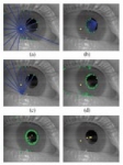
Apr 18, 2006
Cogain project: Communication by Gaze Interaction
The EU-funded five-year project COGAIN (Communication by Gaze Interaction) will attempt to make eye-tracking technologies more affordable for people with disabilities and extend the potential use of the devices to enable users to live more independently.
From the project website:
COGAIN is a network of excellence on Communication by Gaze Interaction, supported by the European Commission's IST 6th framework program. COGAIN integrates cutting-edge expertise on interface technologies for the benefit of users with disabilities. The network aims to gather Europe's leading expertise in eye tracking integration with computers in a research project on assistive technologies for citizens with motor impairments. Through the integration of research activities, the network will develop new technologies and systems, improve existing gaze-based interaction techniques, and facilitate the implementation of systems for everyday communication.
Read the full report
20:30 Posted in Research tools | Permalink | Comments (0) | Tags: Positive Technology
High Speed, Light-based Brain Activity Detector
From Neuromarketing
Neuroscientists Gabriele Gratton and Monica Fabiani at the University of Illinois Beckman Institute’s Cognitive Neuroimaging Laboratory are using very intense near-infrared illumination to measure neuronal activity in the cortex:
The EROS is a new non-invasive brain imaging method that we are developing at the CNL. Our research has determined that this technique possesses a unique combination of spatial and temporal resolution. This makes it possible to use EROS to measure the activity in localized cortical areas. For this reason, EROS can be used to analyze the relative timing of activity in different areas, to study the order of recruitment of different cortical areas, and to examine the connections between areas. These are all questions that are difficult to study with other brain imaging methods.
According to these researchers, the EROS system can measure very short intervals of activity, down to the millisecond level. Its biggest shortcoming is the inability to detect activity more than a few centimeters deep, but it is a relative unexpensive technique (as compared to fMRI and PET) that is not invasive to the test subject.
More information about EROS can be found in this paper entitled: "Fast and Localized Event-Related Optical Signals (EROS) in the Human Occipital Cortex: Comparisons with the Visual Evoked Potential and fMRI" (Neuroimage 6, 168–180 (1997)
11:05 Posted in Research tools | Permalink | Comments (0) | Tags: Positive Technology
Apr 09, 2006
VRoot: OpenEyes
Via VRoot
 openEyes is an open-source open-hardware toolkit for low-cost real-time eye tracking.
Given the increasing demands for more intuitive computer interfaces, tracking the eye movements of users, which precisely indicates users’ attention states, provides researchers and usability experts with invaluable data. However, the high cost of eye-tracking hardware and the lack of available software that implements long-established eye-tracking methods prohibit many interface developers and researchers from accessing or utilizing critical eye-movement data. In response to the need for more widely accessible eye-tracking hardware and software, Derrick Parkhurst, assistant professor of psychology and associate director of VRAC, created the first open source toolkit for low-cost eye tracking, known as openEyes.
20:39 Posted in Research tools | Permalink | Comments (0) | Tags: Positive Technology