


Nov 06, 2006
Dopamine used to prompt nerve tissue to regrow
Via Medgadget

Georgia Tech/Emory researchers are testing how to use dopamine to design polymer that could help damaged nerves reconnect. Their discovery might lead to the development of new therapies for a range of central and peripheral nervous system disorders:
The discovery is the first step toward the eventual goal of implanting the new polymer into patients suffering from neurological disorders, such as Alzheimer's, Parkinson's or epilepsy, to help repair damaged nerves. The findings were published online the week of Oct. 30 in the Proceedings of the National Academy of Sciences (PNAS).
"We showed that you could use a neurotransmitter as a building block of a polymer," said Wang. "Once integrated into the polymer, the transmitter can still elicit a specific response from nerve tissues."
The "designer" polymer was recognized by the neurons when used on a small piece of nerve tissue and stimulated extensive neural growth. The implanted polymer didn't cause any tissue scarring or nerve degeneration, allowing the nerve to grow in a hostile environment post injury.
When ready for clinical use, the polymer would be implanted at the damaged site to promote nerve regeneration. As the nerve tissue reforms, the polymer degrades.
Wang's team found that dopamine's structure, which contains two hydroxyl groups, is vital for the material's neuroactivity. Removing even one group caused a complete loss of the biological activity. They also determined that dopamine was more effective at differentiating nerve cells than the two most popular materials for culturing nerves -- polylysine and laminin. This ability means that the material with dopamine may have a better chance to successfully repair damaged nerves.
The success of dopamine has encouraged the team to set its sights on other neurotransmitters.
"Dopamine was a good starting point, but we are looking into other neurotransmitters as well," Wang said.
The team's next step is to verify findings that the material stimulates the reformation of synapses in addition to regrowth.
23:14 Posted in Neurotechnology & neuroinformatics | Permalink | Comments (0) | Tags: neurotechnology
Oct 26, 2006
Neurotechnology Industry Organization launched
Zack Lynch (Brainwaves) has announced the launch of the Neurotechnology Industry Organization:
The Neurotechnology Industry Organization (NIO) is “a non-profit trade association that represents a broad spectrum of companies involved in neurotechnology (drugs, devices and diagnostics), neuroscience research centers and brain disease advocacy groups across the United States and the world. NIO’s mission is to accelerate cures for brain and nervous system diseases by promoting the neurotechnology industry’s progress, advocating the industry’s position to government officials, and providing business development services to its members”
PT wishes you good luck for your organization, Zack!
18:39 Posted in Neurotechnology & neuroinformatics | Permalink | Comments (0) | Tags: neurotechnology
Sep 10, 2006
Nanowires Listen In on Neurons
Via Neuroguy
MIT’s Technology Review has an interesting article that describes the development of silicon nanowires to measure small electrical signals on the same neuron:
The research group, led by Charles Lieber, professor of chemistry at Harvard University, has developed techniques for synthesizing large arrays of silicon nanowires, which act as transistors, amplifying very small electrical signals from as many as 50 places on a single neuron. In contrast, the most precise existing methods can pick up only one or two signals from a neuron. By detecting electrical activity in many places along a neuron, the researchers can watch how it processes and acts on incoming signals from other cells.The nanowires are about the same size as the branches that neurons use to communicate with one another. William Ditto, professor of biomedical engineering at the University of Florida, says neurons probably send the same kinds of signals to the nanowires as they do to other neurons. As a result, the nanowires could provide a realistic view of a neuron’s complex firing patterns.
21:21 Posted in Neurotechnology & neuroinformatics, Research tools | Permalink | Comments (0) | Tags: neurotechnology
Aug 04, 2006
Whole-brain connectivity diagrams
Via BrainTechSci
In a previous post I covered BrainMaps, the interactive zoomable high-resolution diagram for whole-brain connectivity. Now the project provides a powerful new feature, the interactive visualization of brain connectivity in 3D.
From the download page:
Welcome to nodes3D, a 3D graph visualization program written by Issac Trotts in consultation with Shawn Mikula, in the labs of Edward G. Jones. On startup, nodes3d will download a graph of gross neuroanatomical connectivity from the MySQL database at brainmaps.org. Future versions will probably support loading of graphs from files or other databases
12:50 Posted in Information visualization, Research tools | Permalink | Comments (0) | Tags: neurotechnology
Jul 18, 2006
Second Geoethical Nanotechnology workshop
Re-blogged from KurzweilAI.net
The Terasem Movement announced today that its Second Geoethical Nanotechnology workshop will be held July 20, 2006 in Lincoln, Vermont. The public is invited to participate via conference call.The workshop will explore the ethics of neuronanotechnology and future mind-machine interfaces, including preservation of consciousness, implications for a future in which human and digital species merge, and dispersion of consciousness to the cosmos, featuring leading scientists and other experts in these areas.
The workshop proceedings are open to the public via real-time conference call and will be archived online for free public access. The public is invited to call a toll-free conference-call dial-in line from 9:00 a.m. - 6:00 p.m. ET. Callers from the continental US and Canada can dial 1-800-967-7135; other countries: (00+1) 719-457-2626.
Each workshop presentation is designed for a 15-20 minute delivery, followed by a 20 minute formal question and answer period, during which time questions from the worldwide audience will be invited. Presentations will also be available on the workshop's website
00:05 Posted in Brain training & cognitive enhancement, Brain-computer interface, Neurotechnology & neuroinformatics | Permalink | Comments (0) | Tags: neurotechnology
Jul 06, 2006
Recording of electrical activity by a multi-transistor array (MTA)
M. Hutzler, A. Lambacher, B. Eversmann, M. Jenkner, R. Thewes, and P. Fromherz
Journal of Neuropyhsiology. Preprint online (May 10, 2006).
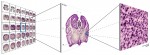
We report on the recording of electrical activity in cultured hippocampal slices by a multi-transistor array (MTA) with 16384 elements. Time-resolved imaging is achieved with a resolution of 7.8 µm on an area of 1 mm2 at 2 kHz. A read-out of fewer elements allows an enhanced time resolution. Individual transistor signals are caused by local evoked field potentials. They agree with micropipette measurements in amplitude and shape. The spatial continuity of the records provides time-resolved images of evoked field potentials and allows the detection of functional correlations over large distances. As examples, fast propagating waves of presynaptic action potentials are recorded as well as patterns of excitatory postsynaptic potentials across and along cornu ammonis.
00:10 Posted in Neurotechnology & neuroinformatics | Permalink | Comments (0) | Tags: neurotechnology