


Nov 26, 2005
EMG Activity in Selected Target Muscles During Imagery Rising on Tiptoes in Healthy Adults and Poststroke Hemiparetic Patients
Authors: Dickstein R, Gazit-Grunwald M, Plax M, Dunsky A, Marcovitz E
The authors sought to gain further knowledge about activation of target muscles during imagery engagement in a motor task. Six hemiparetic patients and 9 healthy participants performed 3 real rises on tiptoes and then, after pausing, 3 imagery rises on tiptoes. Metronome beats guided the rate of rises and descents. Electromyographic (EMG) activity from the medial gastrocnemius and the rectus femoris muscles were monitored bilaterally throughout the performance of both tasks. In 3 healthy participants and 3 individuals with hemiparesis, EMG activity was related to the imagery task in at least 1 of the target muscles. Conversely, in the other participants, motor imagery practice was not accompanied by task-related EMG activity in the monitored muscles. In all cases, the increment in activation level during motor imagery practice was very low in comparison with that of real performance. The findings were not unequivocal; therefore, EMG activity may sometimes, but not always, be recorded during motor imagery practice both in healthy individuals and in poststroke hemiparetic participants. Further research is needed to align motor imagery practice with the objectives of motor rehabilitation.
11:55 Posted in Mental practice & mental simulation | Permalink | Comments (0) | Tags: Positive Technology, mental practice, motor imagery
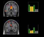
Nov 03, 2005
Mirror therapy for alleviating chronic pain
via Medgadget
McCabe and co-workers from the University of Bath and the Royal National Hospital for Rheumatic Diseases (RNHRD)have published in the journal Clinical Medicine the results of a study, which has investigated the use of mirror as a therapeutic mean to alleviate pain. The treatment consists in asking patients patients suffering from complex regional pain syndrome to carry out routine exercises in front of a mirror.
Results showed that more than half experienced pain relief during and after the exercise and further investigations indicated that even greater improvements can be achieved if the tasks are practiced beforehand.
McCabe explain these findings with the ‘cortical’ model of pain. According to this theory, the brain’s image of the body can become faulty, resulting in a mismatch between the brain’s movement control systems and its sensory systems, causing a person to experience pain when they move a particular hand, foot or limb.
Mirror therapy has proven effective also in the treatment of post-stroke hemiplegia as well as in the rehabilitation of "phantom limb" and visual hemineglect.
More to explore
Sathian K, Greenspan AI, Wolf SL. Doing it with mirrors: a case study of a novel approach to neurorehabilitation.
Neurorehabil Neural Repair. 2000;14(1):73-6.
| Ramachandran VS, Altschuler EL, Stone L, Al-Aboudi M, Schwartz E, Siva N. |
Can mirrors alleviate visual hemineglect? Med Hypotheses. 1999 Apr;52(4):303-5.
Altschuler EL, Wisdom SB, Stone L, Foster C, Galasko D, Llewellyn DM, Ramachandran VS.
Rehabilitation of hemiparesis after stroke with a mirror. Lancet. 1999 Jun 12;353(9169):2035-6.
00:45 Posted in Mental practice & mental simulation | Permalink | Comments (0) | Tags: Positive Technology, mental practice, motor imagery
Oct 26, 2005
Theta brainwave triggered training boosts old brains
It has long been known that hippocampus is involved in the learning process. A study published on the Proceedings of the National Academy of Science (USA) has shown that theta brainwave triggered training can enhance learning rate in old rabbits, contrasting the deficit determined by the aging process. This study could have important implications in the development of nonpharmacological treatments for age-related memory deficits.
Nonpharmacological amelioration of age-related learning deficits: The impact of hippocampal  -triggered training
 rhythm, is known to enhance hippocampal plasticity and accelerate learning rate in young subjects, suggesting that manipulations of activity might be used as a means to counteract impairments related to the aging process. Here, young and older rabbits were given eyeblink conditioning trials either when exhibiting hippocampal (+) or regardless of hippocampal activity (yoked control). Although, as expected, older-yoked control animals showed a learning deficit, the older + group learned as fast as young controls, demonstrating that aging deficits, at least in eyeblink classical conditioning, can be overcome by giving trials during episodes of hippocampal activity. The use of several learning criteria showed that the benefits of hippocampal -triggered training in both age groups during the early phase of acquisition, the enhancement persisted in older animals, peaking during later performance. These findings have implications for theories of age-related memory deficits and may contribute to the development of beneficial treatments. depend on different cognitive or motor processes. Whereas there was a benefit of
16:35 Posted in Mental practice & mental simulation | Permalink | Comments (0) | Tags: Positive Technology, mental practice, motor imagery
Oct 22, 2005
Mental rotation in a patient with an implanted electrode grid in the motor cortex
Neuroreport. 2005 Nov 7;16(16):1795-1800
Authors: Tomasino B, Budai R, Mondani M, Skrap M, Rumiati RI
We investigated the effects of cortical stimulation on mental rotation tasks in a patient with an electrode array placed over his left primary motor cortex. The array was implanted to relieve chronic pain resulting from right brachial plexus damage. Tasks involving motor imagery were slowed down by cortical stimulation, whereas those involving visual imagery were not. When the patient performed the motor-imagery task, the interference effect on response times disappeared if the stimulator was switched off. We also probed two of the sites (anterior-lateral and posterior-medial position), and found that stimulation of the more anterior-lateral one consistently disrupted motor imagery.
19:15 Posted in Mental practice & mental simulation | Permalink | Comments (0) | Tags: Positive Technology, mental practice, motor imagery
Sep 08, 2005
Imagery-induced Cortical Excitability Changes in Stroke
Results of this focal transcranial magnetic stimulation study recently published on Cerebral Cortex by a team of Italian neuroscientists suggest that motor imagery could induce a pronounced motor output enhancement in the hemisphere affected by stroke. These findings strongly support the use of motor imagery as a therapeutic mean in post-stroke motor rehabilitation.
Cicinelli P, Marconi B, Zaccagnini M, Pasqualetti P, Filippi MM, Rossini PM.
Cerebral Cortex, 2005 May 4 (Epub ahead of print)
Focal transcranial magnetic stimulation (TMS) was employed in a population of hemiparetic stroke patients in a post-acute stage to map out the abductor digiti minimi (ADM) muscle cortical representation of the affected (AH) and unaffected (UH) hemisphere at rest, during motor imagery and during voluntary contraction. Imagery induced an enhancement of the ADM map area and volume in both hemispheres in a way which partly corrected the abnormal asymmetry between AH and UH motor output seen in rest condition. The voluntary contraction was the task provoking maximal facilitation in the UH, whereas a similar degree of facilitation was obtained during voluntary contraction and motor imagery in the AH. We argued that motor imagery could induce a pronounced motor output enhancement in the hemisphere affected by stroke. Further, we demonstrated that imagery-induced excitability changes were specific for the muscle 'prime mover' for the imagined movement, while no differences were observed with respect to the stroke lesion locations. Present findings demonstrated that motor imagery significantly enhanced the cortical excitability of the hemisphere affected by stroke in a post-acute stage. Further studies are needed to correlate these cortical excitability changes with short-term plasticity therefore prompting motor imagery as a 'cortical reservoir' in post-stroke motor rehabilitation.
12:45 Posted in Mental practice & mental simulation | Permalink | Comments (0) | Tags: Positive Technology, mental practice, motor imagery
Feb 10, 2005
Brain-imaging study on action observation and acquired motor skills
Earlier studies with monkeys revealed that brain cells called “mirror neurons” respond both when we do something, like pick up an object, and when we simply watch someone else do it. It was known that these neurons fire when we perform an action, but it was unexpected that the same cells also fired when we only saw that action being performed. This new study by Calvo-Merino and coll., recently published on the journal “Cerebral Cortex”, goes a step further by showing that such a system operates differently depending on what a person is physically expert at doing.
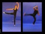
According to the authors of the experiment, this is the first proof that our personal motor repertoire - the things that we have learned to do - changes the way that our brain responds when we see the movement. In particular, these findings suggest that once the brain has learned a skill, it may simulate the skill without even moving, through simple observation: an injured dancer might be able to maintain his skill despite being temporarily unable to move, simply by watching others dance.
These results provide further support to the I-Learning Project clinical rationale, and justify the use of I-Learning technology to better rehabilitate people whose motor skills were damaged by stroke.
Reference: B. Calvo-Merino, D.E. Glaser, J. Grezes, R.E. Passingham and P. Haggard, Action Observation and Acquired Motor Skills: An fMRI Study with Expert Dancers, Cerebral Cortex, Dec. 2004
13:20 Posted in Mental practice & mental simulation | Permalink | Comments (0) | Tags: mental practice, motor imagery